Dental X-rays: which image helps when
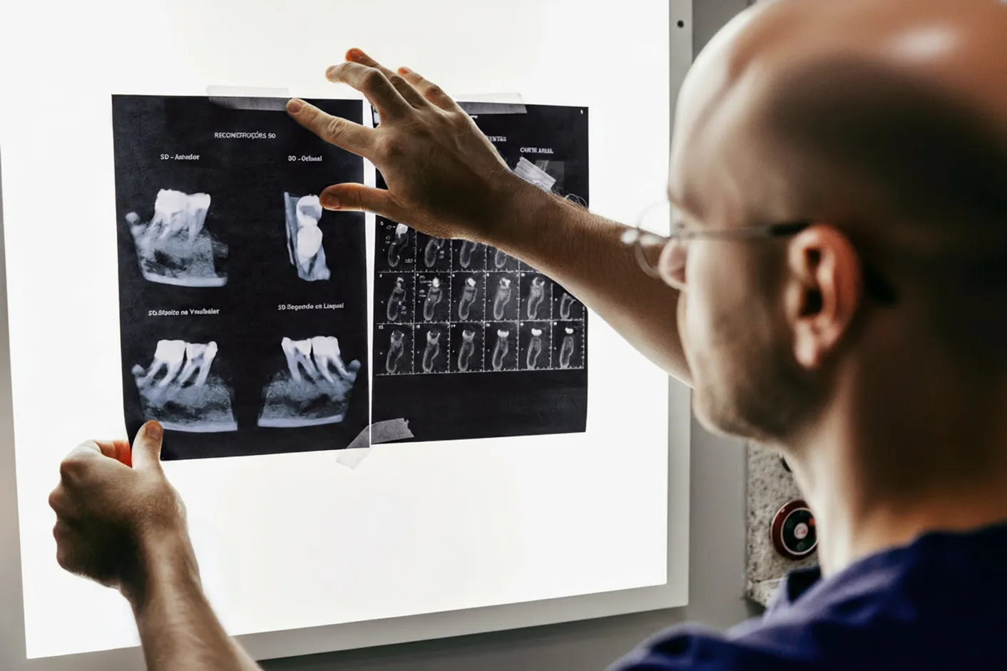
A tooth may look unremarkable in the mouth even though there is an unanswered question about a root, a gap between teeth or the bone. X-rays can provide additional information. However, they are not automatically part of every visit. It must first be established what information is needed and whether it will affect further treatment. The German Radiation Protection Act requires a justifying indication: the expected benefit of each individual application must outweigh the radiation risk. The appropriate image is therefore selected on the basis of the medical history, examination and existing findings.
The short answer
An X-ray requires a specific diagnostic reason. The type of image and its timing are chosen on the basis of the examination and existing findings.
- Why questions and an examination come first
- Pain, an injury, abnormal findings or treatment planning can raise different diagnostic questions. The image should be appropriate to the question concerned. In January 2026, the ADA updated its recommendations on selecting patients for dental X-rays. It emphasises reviewing the medical history, previous images and current examination findings before taking a new image. These US recommendations provide professional information but do not replace the requirements applicable in Germany.
- When a three-dimensional image may be appropriate
- Digital volume tomography, or DVT for short, uses X-rays to create three-dimensional data. The US Food and Drug Administration (FDA) describes specific diagnostic applications for this and emphasises that the examination must be justified. Radiation exposure may be higher than with conventional dental images; the exact extent depends on the equipment and protocol. An examination tailored to the question concerned is particularly important in children.
- Radiation protection: as little as necessary, as much as diagnostically required
- The Radiation Protection Act requires exposure to be limited to the extent necessary for the medical question concerned. Protection begins with avoiding unnecessary images and using appropriate technology. The German Federal Office for Radiation Protection provides an overview of diagnostic applications of radiation. Exposure depends on the type of image and how it is carried out; a single general figure for ‘dental X-rays’ would be misleading.
- Discuss pregnancy and children in advance
- Tell the dental team about an existing or possible pregnancy before the image is taken. This means neither a blanket ban on every necessary examination nor approval without careful consideration. The specific question, possible alternatives and timing must be discussed. Therefore, do not conceal acute symptoms for fear of having an X-ray. Ask what the image can clarify and how the procedure will be adapted.
Why questions and an examination come first
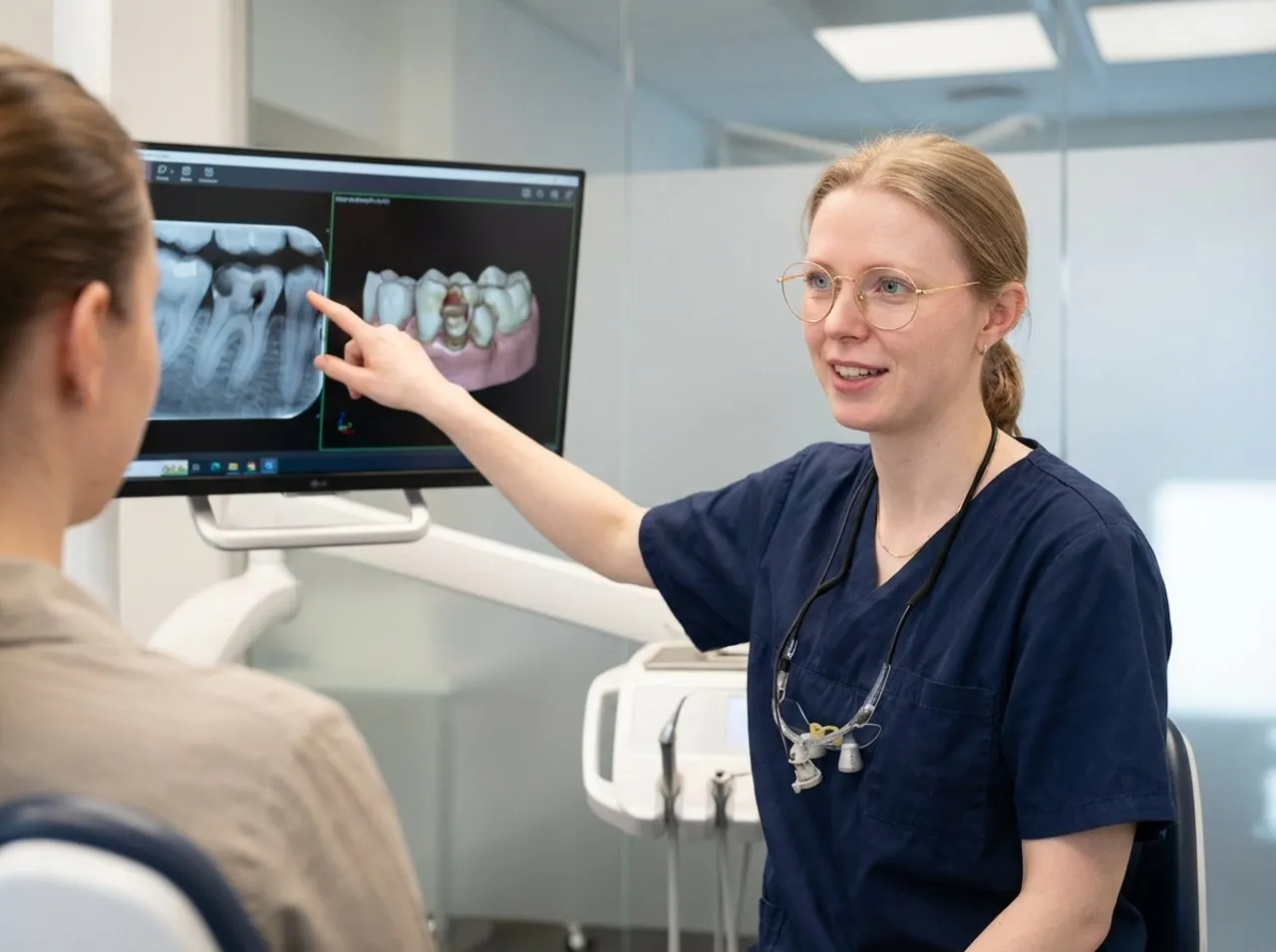
Pain, an injury, abnormal findings or treatment planning can raise different diagnostic questions. The image should be appropriate to the question concerned. In January 2026, the ADA updated its recommendations on selecting patients for dental X-rays. It emphasises reviewing the medical history, previous images and current examination findings before taking a new image. These US recommendations provide professional information but do not replace the requirements applicable in Germany.
You should therefore bring information about recently taken images. Ask whether the practice can request your previous images and whether they are still useful for the current question. This will not prevent every repeat image: an older image may not adequately show the required region or current condition. Conversely, a new image is not necessary simply because a certain period has elapsed. A rigid annual X-ray schedule is not suitable for everyone.
Dental film, bitewing and panoramic image
A small dental film shows a limited area. Depending on the question, the tooth, root and surrounding bone can be assessed. Bitewing images are used in particular to show the crowns of adjacent teeth and can help with the assessment of tooth decay between the teeth . An OPG, also known as a panoramic image, provides a broader overview of the jaws. However, an overview does not automatically replace every detailed image.
| Image type | Basic purpose | Important limitation |
|---|---|---|
| Dental film | Targeted image of a limited tooth/root area. | No overall view of all jaw structures. |
| Bitewing | Image of adjacent tooth crowns and their contact areas. | Not every root or change can be assessed with this. |
| OPG | Overview of larger parts of the teeth and jaws. | A different image is needed for some detailed questions. |
| DVT | Three-dimensional image for a justified specific question. | Not a routine improvement to every examination. |
The table describes typical purposes, not an aid to self-diagnosis. Ask what information is missing in your case. With a root canal treatment , for example, different details are important from those in a general overview. The dental team, with the necessary specialist expertise in radiation protection, decides whether an image is needed.

When a three-dimensional image may be appropriate
Digital volume tomography, or DVT for short, uses X-rays to create three-dimensional data. The US Food and Drug Administration (FDA) describes specific diagnostic applications for this and emphasises that the examination must be justified. Radiation exposure may be higher than with conventional dental images; the exact extent depends on the equipment and protocol. An examination tailored to the question concerned is particularly important in children.
More image information does not automatically mean a better outcome for every patient. Ask whether a simpler image can already provide the information needed, which region is to be covered and how the result will affect planning. DVT is also not equivalent to an optical digital dental impression : an intraoral scan captures visible surfaces and does not work on the same principle.
Radiation protection: as little as necessary, as much as diagnostically required
The Radiation Protection Act requires exposure to be limited to the extent necessary for the medical question concerned. Protection begins with avoiding unnecessary images and using appropriate technology. The German Federal Office for Radiation Protection provides an overview of diagnostic applications of radiation. Exposure depends on the type of image and how it is carried out; a single general figure for ‘dental X-rays’ would be misleading.
Recommendations on lead aprons and other protective equipment have also changed. The German Commission on Radiological Protection explains that inappropriate use can impair the examination or even encourage additional exposure. ‘The same apron for every image’ is therefore not a reliable general rule. Ask for an explanation of the current procedure for your examination. US recommendations on shielding cannot simply be adopted as German practice regulations.
Discuss pregnancy and children in advance
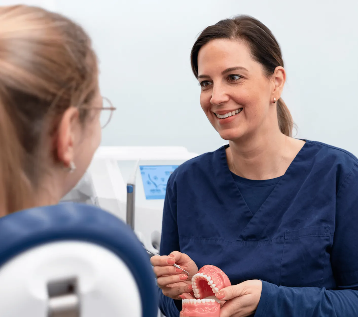
Tell the dental team about an existing or possible pregnancy before the image is taken. This means neither a blanket ban on every necessary examination nor approval without careful consideration. The specific question, possible alternatives and timing must be discussed. Therefore, do not conceal acute symptoms for fear of having an X-ray. Ask what the image can clarify and how the procedure will be adapted.
For children too, the findings and individual needs are what matter. An examination is not automatically ordered simply because of age or a routine check-up. The FDA emphasises the particular attention needed for young patients undergoing DVT. Parents can bring previous images and medical information and ask about the purpose. A professionally justified image must not be equated with an unnecessary one.
These questions can help at your appointment
You do not have to select the imaging technology yourself. However, a clear explanation can make the decision easier to understand. After the image is taken, the findings must be considered together with the symptoms and examination. An unremarkable image does not answer every possible cause of pain; therefore, continue to report any ongoing symptoms and ask about the next step.
Frequently asked questions
No. A new image requires a specific reason. Symptoms, the examination, risk and existing images influence the decision; a general fixed annual schedule does not apply to everyone.
An OPG provides a broader overview of the jaws. A dental film shows a limited area for a specific question. An overview image does not automatically replace every detailed image.
A three-dimensional image can answer specific questions but is not required in every situation. Benefit and radiation exposure are weighed up; more data alone is not a reason for the examination.
A possible or existing pregnancy must be disclosed beforehand. The need, alternatives and timing are assessed individually. A blanket ban or blanket approval would be unreliable.
Recommendations take account of the type of image, the technology and possible interference with the image caused by protective equipment. Ask about the current procedure. An older blanket rule or a US recommendation does not replace a case-by-case decision in Germany.
Tell the practice about previous images and ask about having them transferred. If the images adequately answer the current question, this can avoid unnecessary repeats.