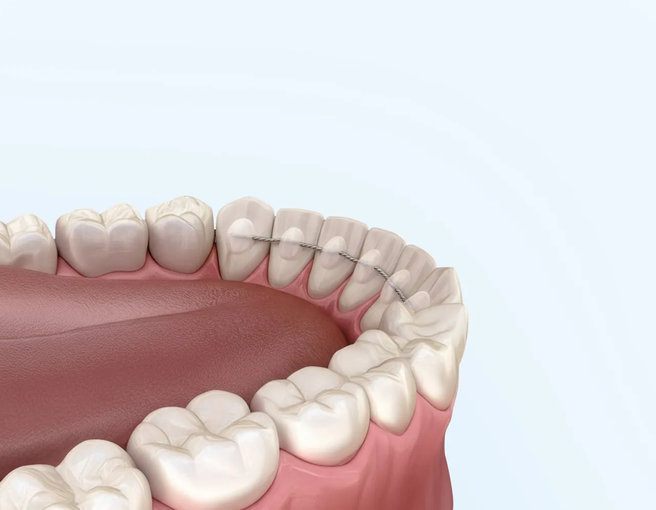
MRI with a retainer or brace: what you should clarify beforehand
You have an MRI scan scheduled, and there is a wire bonded behind your front teeth. Does the retainer need to be removed? A blanket answer would be unreliable. Braces and retainers can have different effects depending on their construction and the examination. Two separate questions are key: is the examination acceptable with the specific appliance, and can the required images be assessed adequately? Therefore, tell the radiology department about your brace when arranging your appointment. Do not remove bonded parts yourself, and do not cancel your MRI appointment simply because of the word ‘metal’.
The short answer
A fixed retainer does not automatically mean that an MRI scan is impossible. The radiology department decides based on the equipment, material and purpose of the examination.
- Safety and image interference are two different issues
- An MRI scanner works with a strong magnetic field and radio waves. The examination team must be informed about metal and implanted devices. Dental fillings and orthodontic appliances are often not a fundamental obstacle, but they can interfere with images of the surrounding area. Such artefacts are distorted or missing image information. A wire that interferes with the images is not automatically dangerous; conversely, suspected minimal image interference is no substitute for a safety check. RadiologyInfo explains the MRI safety check.
- This information helps the radiology department
- Describe what you have: a bonded retainer behind your front teeth, a fixed brace with brackets and a wire, a removable plate or a clear aligner. Specify the upper and lower jaws and mention any additional components on the palate. If you have material information or treatment records, send them or bring them with you as agreed. If the material is unknown, say so openly. Guessing based on colour or trying magnets at home is not a reliable way to identify the material.
- Does a fixed retainer need to be removed?
- Sometimes the appliance can remain in place; sometimes the radiology department requires an adjustment or removal to obtain diagnostically useful images. In a study of 60 people, different brackets and retainers produced different artefact patterns in head and neck images. The region to be assessed was also a determining factor. The individual decision therefore needs to be coordinated between radiology and orthodontics. The original paper examines fixed orthodontic attachments.
- What you should not test yourself
- Do not try to remove the wire with pliers, loosen bonded areas or test it with a magnet. Even a material name alone is not sufficient to assess every component. Simply gather information and leave the decision to the specialists. Store removable appliances safely as instructed. After fixed parts have been temporarily removed, it should be clear what follow-up is planned and who you can contact if the fit changes.
Safety and image interference are two different issues
An MRI scanner works with a strong magnetic field and radio waves. The examination team must be informed about metal and implanted devices. Dental fillings and orthodontic appliances are often not a fundamental obstacle, but they can interfere with images of the surrounding area. Such artefacts are distorted or missing image information. A wire that interferes with the images is not automatically dangerous; conversely, suspected minimal image interference is no substitute for a safety check. RadiologyInfo explains the MRI safety check.
Both questions should therefore be addressed explicitly in a discussion. Do not simply say that your brace is fixed; ask whether it is relevant to the intended scan. A previous examination without problems does not answer this question for every later appointment. The area of the body, scanner type or required image sequences may be different this time. The radiology team responsible makes the decision for the currently planned examination.

This information helps the radiology department
Describe what you have: a bonded retainer behind your front teeth, a fixed brace with brackets and a wire, a removable plate or a clear aligner. Specify the upper and lower jaws and mention any additional components on the palate. If you have material information or treatment records, send them or bring them with you as agreed. If the material is unknown, say so openly. Guessing based on colour or trying magnets at home is not a reliable way to identify the material.
The safety questionnaire is part of the organisational process. Enter your orthodontic appliance on it and also list all other implants and metal objects. Ask if a field does not fit your situation. The team may request additional information. Removable aids and objects are put aside as instructed by the team; bonded parts require separate coordination. RadiologyInfo describes how to prepare for an MRI scan.
Why images of the head and face require special attention
Metal-related image artefacts are particularly significant in the vicinity of the appliance. For images of the mouth, face or brain, this can have different implications than for an examination of a part of the body far away from it. For brain scans, RadiologyInfo explicitly explains that dental materials and braces can affect image quality. Which structures are affected depends on the examination. The fact that a knee MRI was possible cannot therefore simply be assumed to apply to a head MRI. Information about brain MRI.
Scientific studies also show differences between appliances and measurement methods. A study by Sonesson and colleagues compared several orthodontic components at 1.5 and 3 Tesla. The material and imaging technique influenced the artefacts. Such results help with specialist planning, but do not permit a remote diagnosis of your particular brace. You should infer neither reliable clearance nor an automatic requirement for removal from them. The original study explains its design and limitations.

Does a fixed retainer need to be removed?
Sometimes the appliance can remain in place; sometimes the radiology department requires an adjustment or removal to obtain diagnostically useful images. In a study of 60 people, different brackets and retainers produced different artefact patterns in head and neck images. The region to be assessed was also a determining factor. The individual decision therefore needs to be coordinated between radiology and orthodontics. The original paper examines fixed orthodontic attachments.
If removal is required, clarify the timing before anyone detaches the wire: when will the MRI scan take place, who will remove the appliance, and how will the tooth position be maintained afterwards? A retainer is intended to maintain the treatment outcome; its removal should therefore be planned in conjunction with ongoing retention. Also ask about a possible replacement and the associated costs. This does not imply a general waiting period. Particularly for an urgent medical examination, coordination must take place promptly, without postponing the appointment on your own initiative.
What you should not test yourself
Do not try to remove the wire with pliers, loosen bonded areas or test it with a magnet. Even a material name alone is not sufficient to assess every component. Simply gather information and leave the decision to the specialists. Store removable appliances safely as instructed. After fixed parts have been temporarily removed, it should be clear what follow-up is planned and who you can contact if the fit changes.
A message to the radiology department can be brief and clear: I have a fixed retainer in my lower jaw and am due to have an MRI scan of my head. I do not yet have the material information. What documents do you need, and should you liaise with my orthodontic practice? Add the appointment date and your contact details. If two providers give different recommendations, ask them to liaise directly rather than making the decision yourself based on contradictory statements.
A short checklist for the day of the examination
Before setting off, check that the promised documents are available and that any changes to the appliance have actually been made. Take the safety questionnaire with you if you received it in advance. Report any changes since the appointment was booked again. During the examination, there is a way to contact the team; report any unusual warmth, pain or other symptoms immediately. Questions about tooth alignment and retention should then be directed back to your orthodontic team. You can also find basic information about fixed appliances in the guide to fixed braces.
Frequently asked questions
An MRI scan is often possible, but the specific appliance and the planned examination need to be checked. Inform the radiology department in advance; general clearance on the internet is no substitute for its decision.
No. Whether parts can remain in place depends on safety and diagnostic image quality. If removal is necessary, this is coordinated with the orthodontic practice.
Braces can affect images near the mouth and face. Different requirements may apply to brain or facial scans than to an area of the body further away.
Report the missing information and ask the practice treating you for documentation. Describe the position and type of appliance. Do not test the wire yourself with a magnet.
No. Bonded parts should be dealt with professionally. At the same time, clarify how the tooth position will be maintained and monitored after any necessary removal.
It is useful information, but not clearance for every subsequent appointment. The area examined, equipment and imaging technique may be different this time. Declare the appliance again.